Protein Structure and Function
A protein's chain and links support its three-dimensional structure and function. Compare structural proteins, a membrane pore and disulfide links without treating every protein as the same shape.

Structural proteins: collagen and keratin
Protein shape → function
A tendon has to resist a heavy pull. Skin has to survive stretching without tearing. A bacterial outer membrane needs a water-filled pore in the middle of an oily layer. How can one class of molecules — proteins — solve three very different engineering problems?
Collagen · triple helix
Collagen · three-chain triple helix
3D model of collagen showing its triple-helix structure
Follow the three strands. This model shows a molecular building block, not a whole tendon. Many collagen molecules assemble into larger load-bearing structures
| Item | Properties |
|---|---|
| In this model | 3 chains → triple helix |
| At a larger scale | Many molecules → load-bearing fibres |
Collagen · triple helix
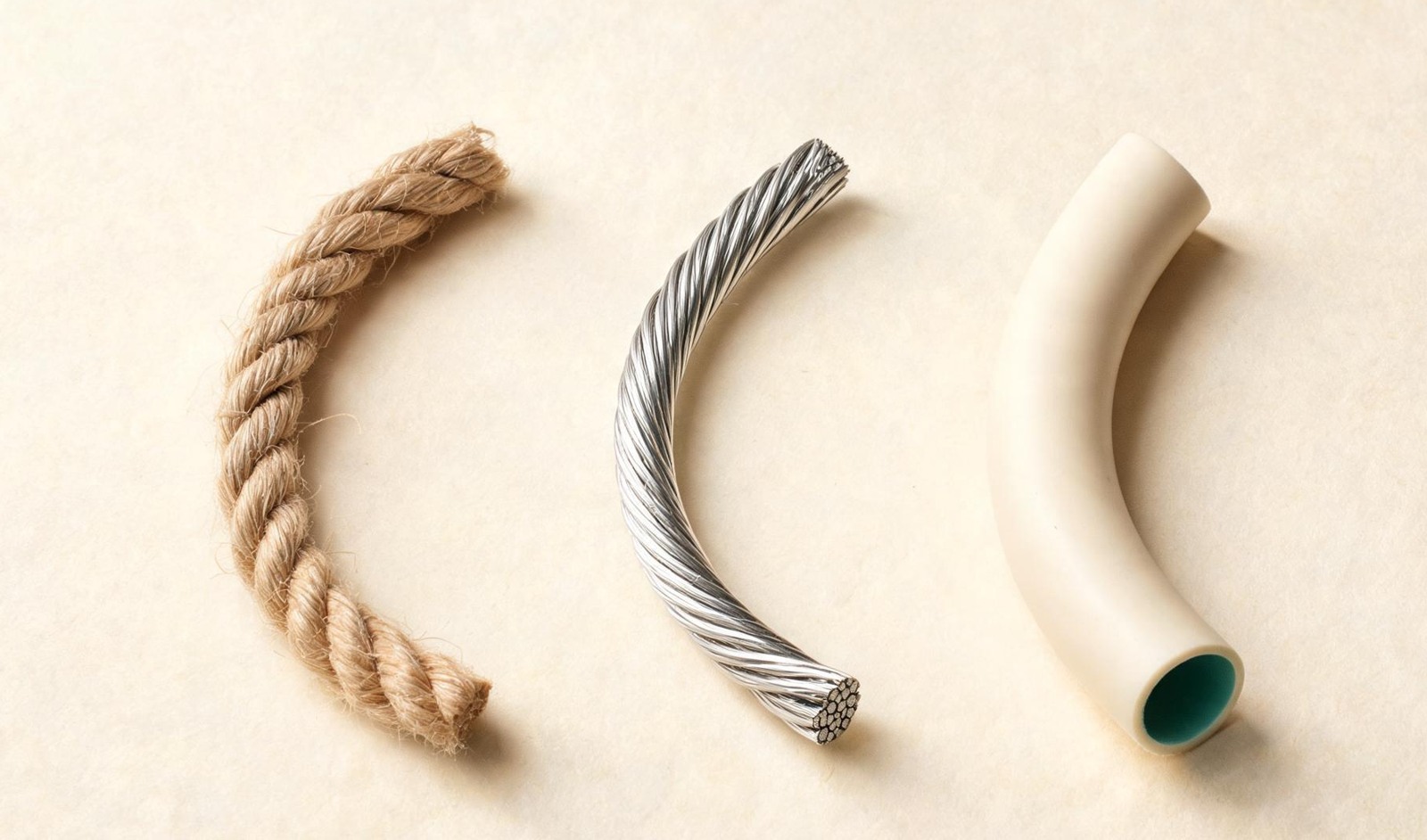
Start with collagen, a structural protein in tendons and skin. Three long polypeptide chains twist together into a rope-like triple helix. In the helical region the sequence follows a Gly-X-Y repeat, so every third residue is glycine — its side chain is just a single hydrogen atom, so it fits into the crowded centre where anything bigger would not
| Item | Properties |
|---|---|
| Triple helix | 3 polypeptide chains |
| Sequence | Gly–X–Y repeats |
| Crowded centre | Gly: small R = H |
Keratin 5 + Keratin 14
Keratin 5 + 14 · coiled-coil dimer
3D model of keratin showing two protein chains coiled together
These are keratins 5 and 14, an epithelial keratin pair found together in cells at the base of the epidermis. As a pair they build coiled-coil dimers, which further assemble into intermediate filaments that help the cells resist mechanical stress
| Item | Properties |
|---|---|
| Epithelial pair | Keratins 5 + 14 |
| Assembly | Dimers → intermediate filaments |
| Mechanical role | Resists cell stress |
Keratin · coiled coil
Keratin uses a different pattern. Two α-helical chains coil around each other. Nonpolar side chains line up along the face where the two helices meet, and hydrophobic attraction holds them together; more polar side chains stay on the outside, where they can meet water
| Item | Properties |
|---|---|
| Coiled coil | 2 α-helical chains |
| Inner interface | Nonpolar faces meet |
| Outer surface | Polar groups meet water |
A pore through a membrane
Porin · 16-strand β-barrel
Porin · a 16-strand β-barrel
3D model of a porin protein with a barrel-shaped structure
Look straight down the barrel. Along a β-strand, side chains point alternately in and out. Nonpolar side chains dominate the outside so the barrel can sit in the membrane, while more water-friendly groups line the pore itself
| Item | Properties |
|---|---|
| Outside | Nonpolar · meets the membrane |
| Inside the pore | Water-friendly groups |
Porin surfaces
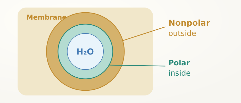
A membrane pore needs almost the opposite arrangement: a nonpolar outer surface that can sit inside the oily membrane, and a water-filled channel down the middle. Porins do this with β-strands curled into a barrel; along many strands, side chains alternate: one points into the membrane, the next points into the water tunnel
Disulfide links stabilise protein structure
Insulin · two chains
Insulin · two chains, three disulfide bonds
3D model of insulin showing its two linked protein chains
Insulin has two chains. Two disulfides connect them; a third lies within chain A. Getting the correct links matters for the hormone's functional structure
| Disulfide links | Number |
|---|---|
| Between chains A and B | 2 |
| Within chain A | 1 |
| Total | 3 |
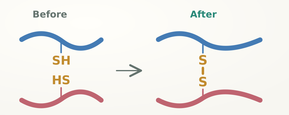
Cysteine → S–S link
Shape can also be locked in with covalent links. When two cysteine side chains are held close together in an oxidising environment, their –SH groups lose hydrogen and join sulfur to sulfur, forming a covalent disulfide bond. This can staple distant parts of one chain, or hold two chains to each other