Cardiac Conduction and AV Node Delay
The cardiac conduction pathway coordinates activation before contraction. Follow the initial signal, atrial spread, AV node delay and ventricular activation to explain the timing of a heartbeat.
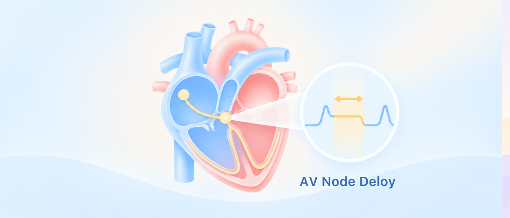
A coordinated heart
A coordinated team
3D heart model showing the cardiac conduction system in context
A coordinated team
The atria are the upper chambers that receive blood. The ventricles are the lower chambers that pump it onward
| Item | Properties |
|---|---|
| Atria | Upper chambers; receive blood |
| Ventricles | Lower chambers; pump blood |
Activation order
Start, spread and delay
Atria
3D heart model highlighting the atria in the conduction sequence
Atria
Most ventricular filling happens while the ventricles relax. The atria then squeeze in a final amount of blood
| Item | Properties |
|---|---|
| Relaxation | Most filling |
| Atrial squeeze | Final filling contribution |
1. Start: The S A node in the right atrium normally starts the electrical signal
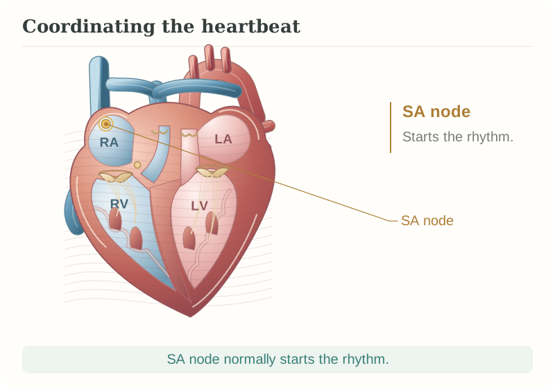
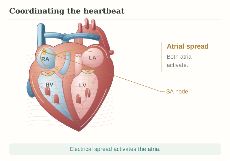
2. Spread: The signal spreads across the atria and triggers their contraction
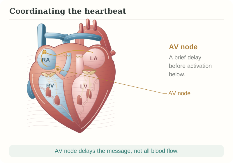
3. Delay: The A V node briefly slows the signal, giving the atria time to finish their contribution
Activate, then contract
Ventricles
3D heart model highlighting the ventricles in the conduction sequence
Ventricles
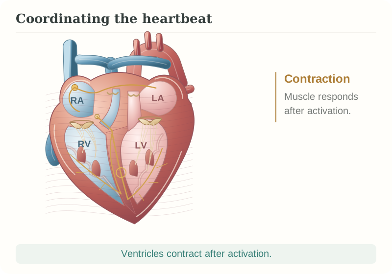
1. Activate: Conducting pathways carry the signal into the ventricles
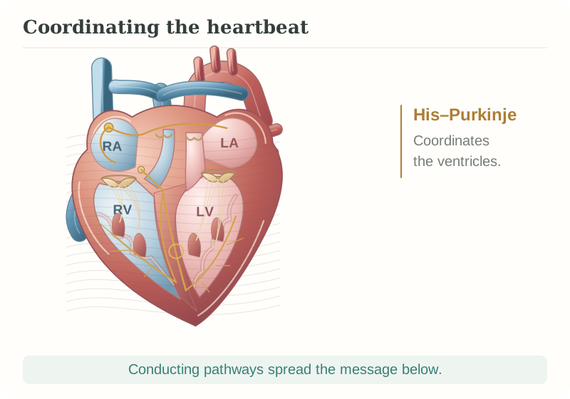
2. Contract: The ventricular muscle contracts after the signal arrives