Proteins in Everyday Life
Protein structure supports different jobs in tissues and cells. Compare collagen, elastin and cartilage with haemoglobin's oxygen transport and a protease's action on peptide bonds.

Skin: strength and recoil
Skin · under the surface
Skin · epidermis and dermis
3D model of skin tissue with a collagen-rich layer beneath the surface
This teaching model shows a slice of human skin. Below the epidermis, the dermis contains a network of collagen fibres that strongly resists pulling, together with elastin and other matrix components
| Item | Properties |
|---|---|
| Dermis | Below the epidermis |
| Fibre network | Collagen + elastin |
Collagen + elastin
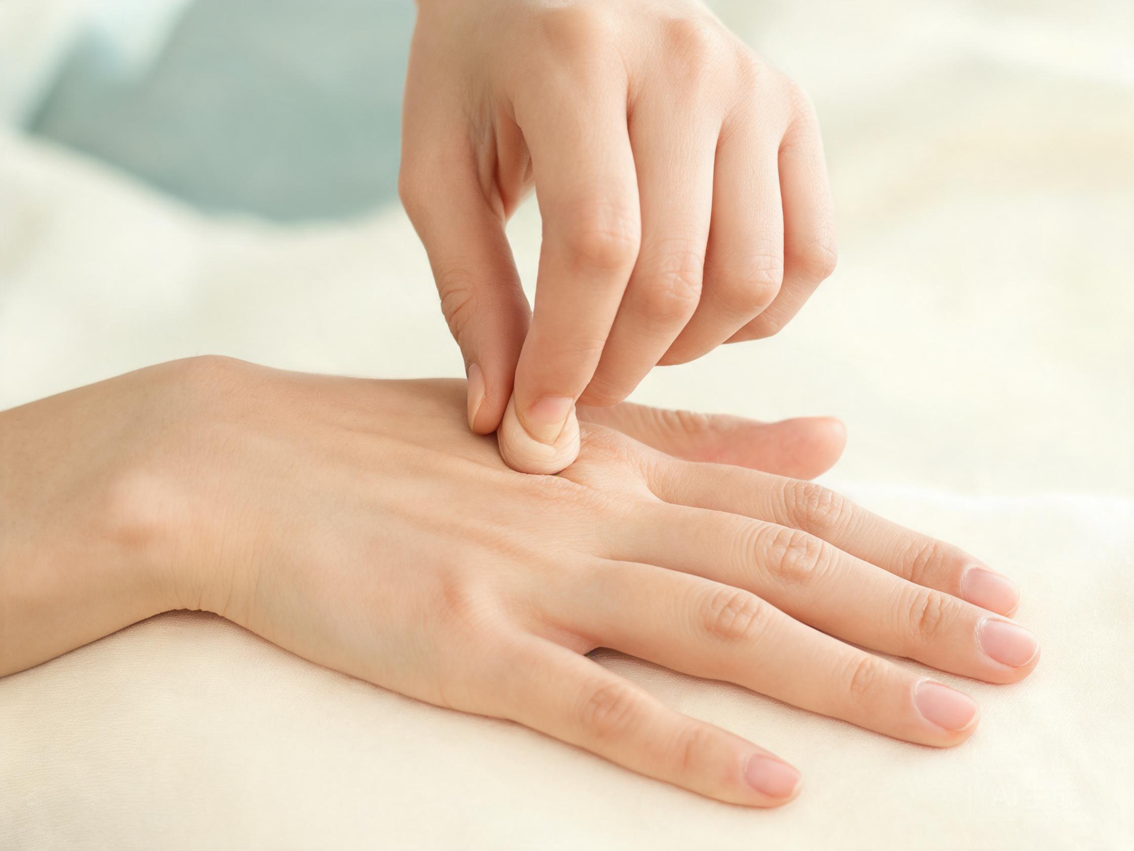
Gently pinch the back of your hand and let go. Your skin resists the pull, and then springs back. Inside the dermis, collagen fibres supply the tensile strength, and elastin fibres give the recoil. Skin is not one protein; it is a composite tissue built around a protein-rich network
| Item | Properties |
|---|---|
| Collagen | Resists pulling |
| Elastin | Springs back |
Protein job 1 · structure
Cartilage: resist compression
Cartilage · a hydrated cushion
Articular cartilage · a hydrated composite
3D model of cartilage tissue containing collagen and water
This is a knee model. The highlighted teal region is articular cartilage. Collagen provides a strong framework, while proteoglycans attract water; together they make a hydrated material that resists compression
| Item | Properties |
|---|---|
| Collagen | Strong framework |
| Proteoglycans + water | Hydrated material |
| Together | Resist compression |
Cartilage resists compression
Now bend one knee. Between the bones sits a smooth, slightly springy layer of articular cartilage. It resists compression instead of pulling
Between bones: compression resistance
From tissue to amino-acid chains
Collagen · from tissue to molecule
Collagen · three amino-acid chains
3D model of a collagen molecule with a repeating chain structure
At molecular scale, each collagen unit contains three polypeptide chains wound together. Every polypeptide chain is built from smaller units called amino acids; a protein can contain one chain or several
| Item | Properties |
|---|---|
| Collagen molecule | 3 polypeptide chains |
| Each polypeptide | Linked amino-acid residues |
| Protein | One chain or several |
Collagen fibre → molecule
Zoom past the tissue and pull one collagen fibre out. What are we actually holding?
Protein = one or more amino-acid chains
Haemoglobin: reversible oxygen transport
Haemoglobin · four oxygen-binding sites
Haemoglobin · four chains and four haem groups
3D model of hemoglobin made of four protein chains
This model is built from the real experimental structure of haemoglobin, drawn as a folded backbone. Four chains pack together: two crimson alpha subunits and two rose beta subunits. Each chain cradles one flat red haem group, and the gold sphere at the centre of each haem is the iron ion — in its Fe²⁺, ferrous form — where one O₂ molecule binds reversibly, without being oxidised into rust
| Model colour | Represents |
|---|---|
| Crimson | α chain |
| Rose | β chain |
| Red disc | Haem group |
| Gold sphere | Fe²⁺ ion |
Not every protein builds tissue. Take a breath. Most oxygen carried in your blood binds reversibly to haemoglobin inside red blood cells
Blood O₂ binds reversibly to haemoglobin
Reversible O₂ transport
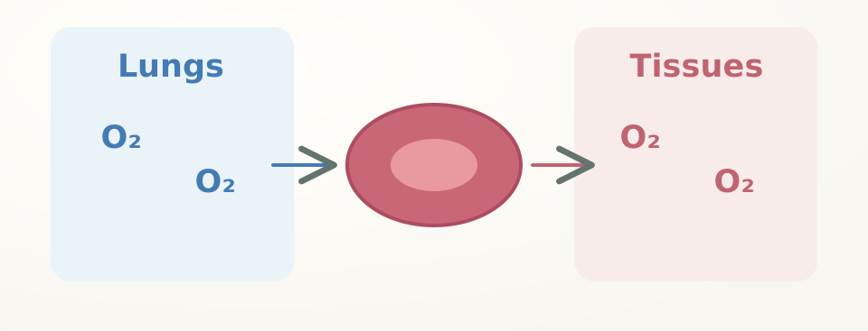
One haemoglobin molecule has four chains and four haem groups, so it can bind up to four oxygen molecules. Binding is reversible: O₂ loads in the lungs and can be released in the tissues
| Item | Properties |
|---|---|
| Binding capacity | 4 haem groups → up to 4 O₂ |
| Reversible transport | Lungs → tissues |
Protease: catalyse peptide-bond hydrolysis
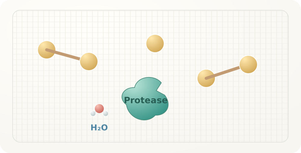
One last stop. Read the back of a laundry detergent bottle. Many list enzymes. Enzymes are proteins that speed up specific reactions
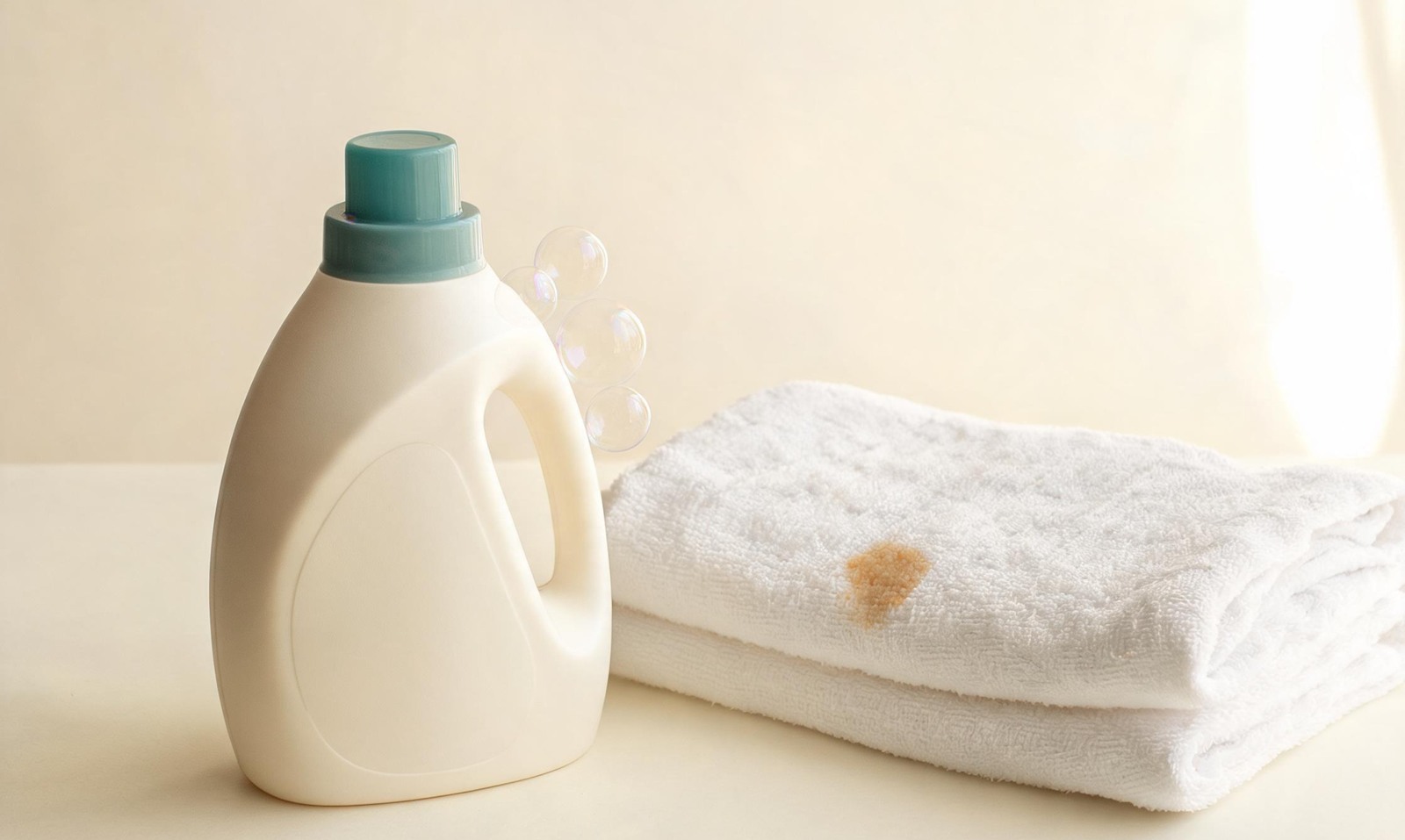
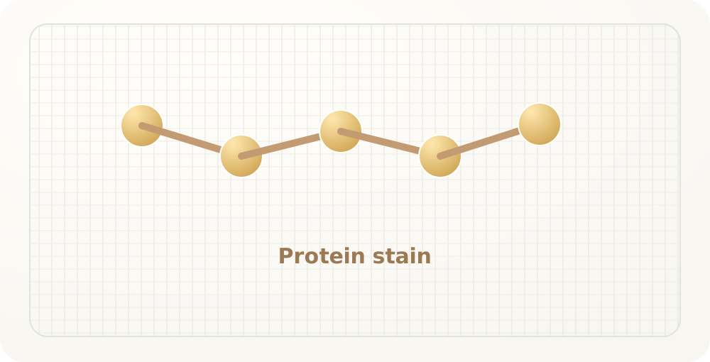
A protease is an enzyme that hydrolyses peptide bonds in proteins. On a blood, milk, or egg stain, it cuts protein molecules into smaller fragments that are easier for the wash to remove